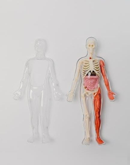
General Overview of the Heart
The heart is a cone-shaped, fist-sized organ located centrally between the lungs, functioning as the circulatory system’s core, pumping blood through its chambers and valves efficiently.
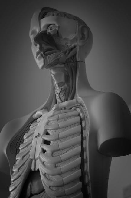
1.1. Position, Size, and Shape of the Heart
The heart is centrally located in the chest cavity, slightly to the left of the midline, nestled between the lungs. It is roughly the size of a clenched fist, weighing approximately 250-350 grams. Its shape is conical, with the apex pointing downward and the base facing upward. This anatomical positioning ensures efficient blood circulation and pulmonary function, while its size and shape optimize its pumping efficiency within the thoracic space.
1.2. Role of the Heart in the Circulatory System
The heart is the central organ of the circulatory system, responsible for pumping blood throughout the body. It ensures oxygenated blood is delivered to tissues and deoxygenated blood is returned to the lungs for oxygenation. By generating blood pressure, the heart maintains continuous circulation, supplying nutrients and oxygen while removing waste products. This vital function sustains cellular metabolism, enabling overall bodily functions and maintaining life.
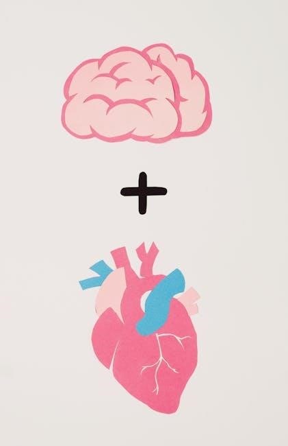
Layers of the Heart Wall
The heart wall consists of three layers: the outer epicardium, the muscular myocardium, and the inner endocardium, each serving unique protective, contractile, and lining functions respectively.
2.1. Epicardium (Outer Layer)
The epicardium is the heart’s outermost layer, forming a protective sac that anchors the heart in the chest cavity. It is a fibrous membrane, part of the pericardium, reducing friction during contractions. Composed of connective tissue, it contains blood vessels and nerves, aiding in the heart’s structural support and function. The epicardium also houses coronary vessels, supplying the heart muscle with oxygen and nutrients, ensuring proper cardiac function and overall cardiovascular health.
2.2. Myocardium (Middle Layer)
The myocardium is the thick, muscular middle layer of the heart wall, primarily responsible for contraction and pumping blood. Composed of cardiac muscle cells, it contains sarcomeres, enabling strong contractions. This layer varies in thickness, with the left ventricle being the thickest due to its high workload. The myocardium is richly supplied with blood vessels and nerve fibers, ensuring efficient oxygenation and coordination of heartbeats. Its thickness and structure allow it to generate the force needed for blood circulation throughout the body.
2.3. Endocardium (Inner Layer)
The endocardium is the thin, smooth innermost layer of the heart, lining the chambers and valves. It prevents friction and ensures smooth blood flow. Composed of endothelial cells, it aids in maintaining the heart’s internal environment. The endocardium also plays a role in regulating cardiac function and preventing thrombosis. Its smooth surface is crucial for efficient blood circulation, while its elasticity accommodates the heart’s constant contractions and relaxations. This layer is vital for maintaining the heart’s structural integrity and function.
Chambers of the Heart
The heart contains four chambers: the right and left atria, and the right and left ventricles. They work together to ensure efficient blood circulation throughout the body.
3.1. Right Atrium and Ventricle
The right atrium receives deoxygenated blood from the body through the superior and inferior vena cava. It then flows through the tricuspid valve into the right ventricle, which pumps blood to the lungs for oxygenation. The right ventricle’s muscular walls generate sufficient force to propel blood through the pulmonary artery, ensuring efficient gas exchange. Together, these chambers play a crucial role in the pulmonary circulation, maintaining the body’s oxygen supply.
3.2. Left Atrium and Ventricle
The left atrium receives oxygen-rich blood from the lungs via the pulmonary veins. This blood flows through the mitral valve into the left ventricle, which is thicker and more muscular than the right ventricle. The left ventricle pumps blood through the aortic valve into the aorta, the largest artery, supplying oxygenated blood to the entire body. This chamber is vital for systemic circulation, ensuring tissues and organs receive the oxygen needed for metabolism and function.
Valves of the Heart
The heart contains four valves ensuring one-way blood flow. Atrioventricular and semilunar valves prevent backflow, maintaining efficient circulation and proper cardiac function throughout the cardiac cycle.
4.1. Atrioventricular Valves
Atrioventricular valves, located between the atria and ventricles, ensure blood flows in one direction. The mitral valve (left) and tricuspid valve (right) have cusps that open and close, preventing backflow. These valves are critical for maintaining efficient blood circulation and proper cardiac function, ensuring blood moves forward through the heart chambers without leakage; Their proper functioning is essential for overall cardiovascular health and maintaining systemic and pulmonary circulation balance.
4.2. Semilunar Valves
Semilunar valves, located at the bases of the pulmonary artery and aorta, regulate blood flow from the ventricles to the circulatory system. The pulmonary valve controls blood flow to the lungs, while the aortic valve directs oxygenated blood to the body. Both valves have three cusps, ensuring unidirectional blood flow and preventing backflow into the ventricles. Their proper functioning is crucial for maintaining efficient blood circulation and overall cardiac performance, ensuring blood reaches its intended destinations without obstruction.
Conducting System of the Heart
The heart’s conducting system includes the SA node, AV node, Bundle of His, and Purkinje fibers, working together to regulate heartbeat timing and synchronize contractions for efficient blood flow.
5.1. Sinoatrial (SA) Node
The SA node, located in the right atrium near the superior vena cava, acts as the heart’s natural pacemaker. It generates electrical impulses at a rate of about 60-100 beats per minute, setting the rhythm for cardiac contractions. This intrinsic pacing ensures a consistent heartbeat, coordinating the atria’s contraction with the ventricles, maintaining synchronized blood flow through the chambers and into the circulatory system.
5.2. Atrioventricular (AV) Node
The AV node, located between the atria and ventricles near the septum, acts as a critical relay station in the heart’s electrical conduction system. It receives impulses from the atria via the SA node and introduces a delay, ensuring ventricular contraction occurs after atrial contraction. This delay optimizes blood filling and pumping efficiency. The AV node also functions as a backup pacemaker if the SA node fails, maintaining a slower but steady heart rhythm when necessary.
5.3. Bundle of His
The Bundle of His is a group of specialized cardiac fibers that transmit electrical impulses from the AV node to the ventricles. Located in the septal wall between the atria and ventricles, it splits into left and right branches, ensuring synchronized contraction of both ventricles. This rapid conduction pathway is essential for maintaining a coordinated heartbeat, enabling the ventricles to pump blood efficiently. Damage to the Bundle of His can disrupt this coordination, leading to arrhythmias or ventricular dysfunction.

5.4. Purkinje Fibers
Purkinje fibers are large, branching cardiac fibers that originate from the Bundle of His and extend into the ventricular walls. They rapidly conduct electrical impulses, ensuring synchronized contraction of the ventricles. These fibers have fewer myofibrils but abundant mitochondria, enabling high-energy transmission. They play a critical role in maintaining a coordinated heartbeat and preventing arrhythmias. Damage to Purkinje fibers can disrupt ventricular rhythm, leading to potentially life-threatening arrhythmias;

Blood Circulation Through the Heart
Blood circulates through the heart via pulmonary and systemic pathways, ensuring oxygenated blood reaches the body and deoxygenated blood is sent to the lungs for oxygenation.
6.1. Pulmonary Circulation
Pulmonary circulation transports deoxygenated blood from the right ventricle through the pulmonary artery to the lungs. Here, gas exchange occurs: oxygen diffuses into the blood, and carbon dioxide is removed. Oxygen-rich blood returns to the left atrium via the pulmonary veins. This system operates at lower pressure than systemic circulation, ensuring efficient oxygenation of blood before it re-enters the systemic loop.
6.2. Systemic Circulation
Systemic circulation delivers oxygen-rich blood from the left ventricle through the aorta to the body’s tissues. Arteries branch into smaller arterioles and capillaries, enabling oxygen and nutrient exchange. Deoxygenated blood collects in venules and veins, returning to the right atrium. This system operates at higher pressure than pulmonary circulation, ensuring blood reaches all body tissues efficiently before returning to the heart for re-oxygenation and the cycle repeats.
6.3. Coronary Circulation
Coronary circulation supplies oxygen-rich blood to the heart muscle itself. The coronary arteries branch from the aorta, delivering blood to the myocardium through a network of capillaries. Deoxygenated blood collects in the coronary veins and returns to the right atrium via the coronary sinus. This circulation ensures the heart muscle receives the oxygen and nutrients needed for continuous contraction, maintaining its function as a pump for the entire circulatory system. Coronary circulation operates independently of systemic and pulmonary pathways.

Cardiac Cycle
The cardiac cycle is the continuous process of heart contractions and relaxations, enabling blood flow through the chambers and into the circulatory system, repeating approximately 70-80 times per minute.
7.1. Phases of the Cardiac Cycle
The cardiac cycle consists of two main phases: systole and diastole. Systole includes atrial contraction and ventricular ejection, while diastole involves ventricular relaxation and filling. These phases ensure efficient blood circulation, maintaining the body’s oxygenation and nutrient delivery. Proper coordination of these phases is crucial for overall cardiac function. The cycle repeats continuously, regulated by the heart’s intrinsic conduction system and external factors like nervous and hormonal controls.
7.2. Regulation of the Cardiac Cycle
The cardiac cycle is regulated by the autonomic nervous system, with the sympathetic nervous system increasing heart rate and contractility during stress, and the parasympathetic nervous system promoting relaxation and reducing heart rate. The sinoatrial node acts as the natural pacemaker, initiating heartbeats. Ion concentrations, such as calcium and potassium, play a crucial role in electrical activity. Hormones like epinephrine also modulate heart activity, ensuring the cardiac cycle adapts to the body’s needs.
Cardiac Output and Stroke Volume
Cardiac output is the volume of blood pumped by the heart per minute. Stroke volume is the blood ejected per heartbeat. Both determine cardiac efficiency and overall circulatory performance.
8.1. Factors Affecting Stroke Volume
Stroke volume is influenced by preload, contractility, and afterload. Preload refers to the initial stretching of the ventricular muscle fibers, determined by venous return. Contractility is the intrinsic strength of the heart muscle, regulated by sympathetic nervous system activity and hormones like epinephrine. Afterload is the resistance the ventricles must overcome to eject blood, primarily due to systemic vascular resistance. These factors collectively determine the efficiency of blood ejection during each cardiac cycle.
8.2. Factors Affecting Cardiac Output
Cardiac output is influenced by heart rate, stroke volume, and overall cardiovascular health. Heart rate is regulated by the autonomic nervous system, with sympathetic stimulation increasing it and parasympathetic activity decreasing it. Stroke volume depends on preload, contractility, and afterload. Hormones like epinephrine enhance contractility, while blood volume and systemic vascular resistance also play roles. These factors collectively determine the heart’s ability to meet the body’s circulatory demands efficiently.
